Liver

Definition
Hepatocellular Carcinoma
The primitive liver cancer forms in the cells of the liver (hepatocytes), in the bile ducts, or in blood vessels.
The primitive liver cancer is different from the cancer which starts somewhere else in the organism which then propagates to the liver (called secondary or metastatic cancer of the liver).
The liver is one of the most voluminous organs of the human body. It is located in the superior part of the abdomen, on the right side, and is protected by the lower ribs.
Two sections, called lobes, divide the liver, right and left, with the latter being the smaller.
The liver fulfils several essential functions for your health:
- It produces enzymes and bile which facilitate digestion.
- It stores energy, vitamins and minerals, and liberates them into the blood when the body is in need.
- It synthesises proteins which contribute to the coagulation of blood in order to stop bleeding in case you cut or injure yourself.
- It removes from the blood all harmful elements, like alcohol and the organism’s waste products.
- It regularises the quantity of certain chemical substances which are naturally present in the body, for example cholesterol.
The liver gets its supply of blood from two sources. The hepatic artery, which transports blood towards the liver, contains a large quantity of oxygen, coming from the lungs and the heart. The blood which comes from the intestines, is rich in nutriments, and arrives at the liver by the portal vein.
Most of the basic liver cancers develop in the liver cells (hepatocytes), and are called hepatocellular carcinomas.
Cholangiocarcinoma, also known as bile duct cancer, is less frequent, and starts life in the bile ducts.
Bile ducts allow the bile produced by the liver to flow to the gallbladder, where it is kept until needed by the organism for the digestive process.
The information contained here concerns hepatocellular cancers, but cholangiocarcinomas are often treated in the same manner.
The secondary liver cancer (also called the metastatic cancer of the liver), comes from a cancer situated elsewhere in the organism, which will then propagate to the liver.
Causes and Risk Factors
The primitive liver cancer cannot be attributed to a single cause, but certain factors together increase the risk:
- A chronic liver infection (h-Hepatitis B or C).
- Cirrhosis: a lesion of the liver caused by a hepatitis, excessive consumption of alcohol over a long period, and certain hereditary factors.
- Exposure to Aflatoxin: a natural chemical product produced by certain moulds developing on nuts and cereals when improperly stored (a phenomenon more frequent in Africa and Asia).
- Exposure to certain industrial chemical products (particularly vinyl chloride and arsenic).
- The long term use of anabolic steroids (hormones which certain athletes take to increase their muscle strength).
- Certain metabolic disorders, such as hemochromatosis (the excessive accumulation of iron in the liver).
The primitive liver cancer may sometimes develop in the absence of all these risk factors.
Diagnosis of the primitive liver cancer
It is probably your GP/Family Doctor who suspected the presence of a primitive liver cancer after having examined you and asked about your health and medical history.
To do this, the doctor would palpate your abdomen and your pelvic area to see if the liver, the spleen and the adjacent organs, present any suspicious lumps or have changed shape or size.
The doctor will also check if there is an abnormal accumulation of liquid in the abdomen (Acsites) and will examine your skin and your eyes to check for signs of jaundice (icterus).
To confirm their diagnosis, the doctor would also make a few analyses, which would also establish the « stage » of the cancer.
It could be that you underwent one or more of the following tests.
Blood analyses:
- Using your blood sample, we can check the quantity and aspect of the different types of blood cells. The results of the analyses will show in what measure your organs function normally and can supply the indications suggesting the presence or not of a cancer.
- A hepatic function test helps to evaluate the way your liver functions. Another test measures the time your blood takes to clot.
- It is also possible to check the presence in your blood of proteins called tumour markers. Liver cancer cells produce a tumour marker called Alpha-fetoprotein (AFP). High levels of AFP may be an indication of a cancer.
Imaging techniques:
These techniques provide and in-depth examination of tissue, organs and bones.
X-rays, ultrasound, a CT scan, a magnetic resonance imaging (MRI) scan and a bone scintigraphy, are a number of ways that your medical team can use to obtain an image of a tumour and check if it is growing. These tests are generally painless and require no anaesthesia.
You could also have a special x-ray called an arteriogram (or angiogram). A particular colourant is injected into an artery in the groin. The colourant infiltrates the blood vessels in the liver, which allows the doctor to see it more clearly.
Biopsy:
A biopsy may be required to establish with certitude the diagnosis of cancer.
This procedure consists of taking a sample of the cells of the organism in order to examine them under a microscope. If the cells are cancerous, the next stage is to determine how rapidly they can multiply.
There are different types of biopsy:
- With a drill biopsy, the doctor inserts the needle into an incision made in the abdomen in order to remove a voluminous fragment of tissue.
- When a fine needle is used it draws in a small quantity of tissue from the abnormal area of the liver.
In both cases, the doctor can use ultrasound imaging or a volume CT scan to place the needle in the correct area.
A local anaesthetic is used to numb the area being examined.
As there is a risk of bleeding following a biopsy of the liver, you may have to stay in the hospital for a few hours, or overnight, after the procedure.
Laparoscopy/Keyhole surgery:
With a laparoscopy, a straight but supple tube with a light and camera at its extremity, is introduced into the abdomen through a small incision. The surgeon can examine the liver and the other internal organs around it and take several small samples of tissue, which are sent for a histological analysis (biopsy).
A laparoscopy can be carried out under a simple local anaesthetic, but at the hospital we habitually use a general anaesthetic.
Analysing the development stage of a primitive cancer of the liver
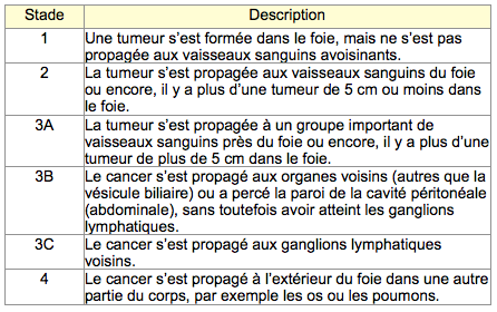
Once the diagnosis of a cancer is confirmed, and your medical care team has obtained all the necessary information, it is essential to determine the development Stage of the cancer.
This process consists of defining the size of the tumour, and checking if it has spread from the site where it started.
Four Stages have been defined for primitive cancer of the liver. Stage 3 is divided into three sub-groups.

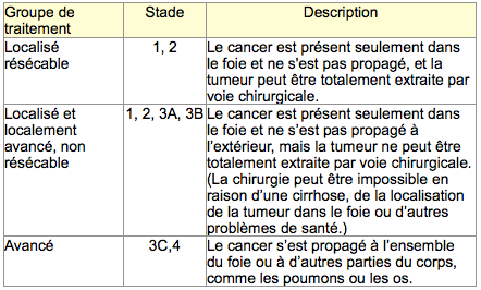
In the case of a primitive cancer of the liver, we can also regroup the different Stages relative to the possible treatments. There are three groups of treatments.

It is important to know the Stage of your cancer, because it is what will help you, and your medical care team, to choose the treatment which suits you the best.
Treatment for primitive liver cancer
The choice of treatment for a primitive cancer of the liver also depends on:
- The condition of the liver.
- The size and location of the tumour, as well as the number of tumours present in the liver.
- The propagation, or not, of the cancer outside the liver.
Riferimenti bibliografici
- Reference 1 : Test1
- Reference 2 : Test
- Reference 3 : Test
- Reference 4 : Test
- Reference 5 : Test
- Reference 6 : Test
- Reference 7 : Test
- Reference 8 : Test
- Reference 9 : Test


