Liver
– – Definition Hépatocellulaire carcinoma
Hépatocellulaire carcinoma
The primitive cancer of the liver is formed in the cells of the liver (hépatocytes), the bile ducts or the blood-vessels.
The primitive cancer of the liver is different from the cancer which occurs elsewhere in the organization to be propagated then with the liver (called secondary or metastatic cancer of the liver).
The liver is one of the bulkiest bodies of the human body. It is placed in the upper part of the abdomen, the right side, and is protected by the lower coasts.
Two sections, called lobes, divide the liver, the left right and it, this last being smallest.
The liver fulfills several functions essential with your health:
- It produces enzymes and bile which facilitate digestion,
- It stores energy, vitamins and minerals and releases them in blood when the body needs some,
- It synthesizes proteins which contribute to the coagulation of blood in order to stop the bleeding in the event of cut or of wound.
- It removes blood from the harmful elements like the alcohol and waste of the organization,
- It regularizes the quantity of certain chemical substances naturally present in the body, for example cholesterol.
The liver is supplied in blood starting from two sources. The hepatic artery, which transports blood towards the liver, contains a great quantity of oxygen, coming from the lungs and the heart. The blood which comes from the intestines, rich in nutrients, arrives towards the liver by the portal vein.
Most primitive cancers of the liver develop in the cells of the liver (hépatocytes), one calls them carcinomata hépatocellulaires.
The cholangiocarcinomist, less frequent, occurs in the bile ducts, which convey the bile produced by the liver to the gall bladder, where it will be preserved until the organization needs for the digestive process.
The contained information here relates to cancers hépatocellulaires, but the cholangiocarcinomists are often treated same manner.
The secondary cancer of the liver (also called metastatic cancer of the liver), comes from a cancer located elsewhere in the organization and which thereafter will be propagated with the liver.
– – Liver causes and risk factors
Causes and Factors
Causes and Risk factors
The primitive cancer of the liver is not ascribable to a single cause, but certain factors seem associated at an increasing risk:
- A chronic hepatic infection (H-Hepatitis B or C)
- Cirrhosis: lesion of the liver caused by hepatitis, the alcohol excessive consumption during one long period and certain hereditary factors.
- The exposure to Aflatoxine: a natural chemical substance produced by the mould developing on walnuts and cereals under bad conditions of storage (more frequent phenomenon in Africa and Asia).
- The exposure to certain industrial chemical products (in particular the vinyl chloride and arsenic).
- The long-term use of steroids anabolics (hormones which certain athletes consume to increase their muscular force).
- Some turbid of the metabolism, such as the hémochromatose (excessive accumulation of iron by the liver).
The primitive cancer of the liver can sometimes develop in the absence of all these risk factors
– – Diagnosis of the primitive cancer of the liver
Diagnosis
Diagnosis of the primitive cancer of the liver
It is probable that your doctor suspected the presence of a primitive cancer of the liver after you of having examined and questioned on your medical health status and your antecedents.
With this intention, the doctor will carry out palpations on the level of your abdomen and your basin to see whether the disastrous liver, it and the neighbouring bodies have suspect masses or changed form or size. The doctor will also check if it there has an abnormal accumulation of liquid in your abdomen (acsite) and examines your skin and your eyes in order to detect signs of jaundice (ictère).
To confirm his diagnosis, the doctor will resort to certain analyses, which will be able to also make it possible to establish the “stage” of cancer. It may be that you have to pass one or more from the following tests.
Blood analyses
- From samples of your blood, one checks the quantity and the appearance of the various types of blood cells. The result of the analyses show your bodies up to what point function normally and can provide indications suggesting the presence or not of a cancer.
- A test of the hepatic function will make it possible to evaluate the operation of your liver. Another test will measure time that puts your blood to coagulate.
- It is also possible that one checks the presence in your protein blood called tumoral markers. The cancer cells of the liver manufacture a tumoral marker of the alpha-fetoprotein name (AFP). High concentrations of AFP can be an indication of cancer.
Techniques of imagery
These techniques make it possible examine a closer examination of fabrics, bodies and bones. Radiography, l’echography, tomodensitometry [TDM], the imagery by magnetic resonance [MRI] and the osseous scintiscanning are as many means for your medical team of obtaining an image of the tumour and of checking if it extended. These tests are generally without pain and do not require any anaesthesia.
You could also pass a special radiography called artériogramme (or angiogramme). One injects initially a particular dye in an artery of the groin; the dye infiltrates then in the blood-vessels of the liver, which makes it possible to the doctor more clearly to see it.
Biopsy
A biopsy can be necessary to establish with certainty a diagnosis of cancer.
This intervention consists in taking cells of the organization in order to examine them under the microscope. If the cells are cancerous, their speed will have then to be determined to multiply. There exist various types of biopsies:
- During a biopsy by drilling, the doctor inserts a needle in an incision practised in the abdomen in order to withdraw a bulky fabric fragment.
- The puncture with the fine needle uses a thin needle to aspire a modest amount of fabric of the abnormal area of the liver.
In both cases, the doctor will be able to resort to the imagery by ultrasounds or the tomodensitometry to place the needle at the good place. A local anaesthetic will be used to anaesthetize the area under examination. As there is a risk of bleeding following a biopsy of the liver, it may be that you must remain at the hospital during a few hours or all the night after the intervention.
Laparoscopy
At the time of a laparoscopy, a narrow and flexible tube, provided with a light and a camera at its end, is introduced by a small incision into the abdomen. The surgeon will examine the liver as well as the other close internal bodies and will take several small samples which will be sent in histological analysis (biopsy).
A laparoscopy can be practised under simple local anaesthesia, but it takes usually place at the hospital under general anaesthesia.
– – Stadification of the primitive cancer of the liver
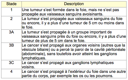
Once the diagnosis of cancer is confirmed and that your medical team collected all the necessary information, it is then necessary to determine the stage of cancer.
The stadification of cancer consists in defining the size of the tumour and checking if it developed beyond the site where it occurred.
Four stages were defined for the primitive cancer of the liver. Stage 3 is divided into three sub-groups.

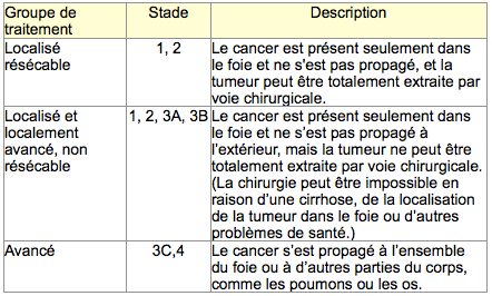
In the case of the primitive cancer of the liver, one can also gather the various stages according to the possible treatment. There exist three groups of treatment.

It is important to know the stage of your cancer, because it is what will help you, you like your medical team, to choose the treatment which is appropriate to you best.
– – Treatment of the primitive cancer of liver (CHC)
The choice of the treatment for a primitive cancer of the liver will also depend:
- condition of the liver;
- size and localization of the tumour, like amongst tumours present in the liver;
- propagation or not of cancer outside the liver.


Commentaires récents